Showing posts with label FOAM. Show all posts

Sunday, November 13, 2016

Slaying Prehistoric Practice - Killing off the Routine Pulse Check

The Gist: It's time to kill off the routine pulse check every two minutes. While some providers and systems have moved away from routine pulse checks, perfunctory cessation in compression to identify pulses happens as 'the norm' across the United States. Perform rhythm checks, which take far less time, taking less than 10 seconds to do so. Pulse checks may be indicated if there is a perfusing rhythm and/or other signs of ROSC. 


Most of us recognize that, to the best of our knowledge, interruptions in chest compression may be deleterious to return of spontaneous circulation and, theoretically, neurologic outcome [2]. We may even devise complicated ways of reducing time without compressions including mechanical compressions and hands-on defibrillation. These interventions are not evidence-based and the yield is minimal. A more simple fix exists - stop checking for pulses routinely.  This isn't some novel wild idea.

The American Heart Association guidelines do not recommend any specific interval for pulse check after the first initial pulse check for healthcare providers [3].  Beginning in 2010,  however, the AHA guidelines begin discouraging routine pulse checks:
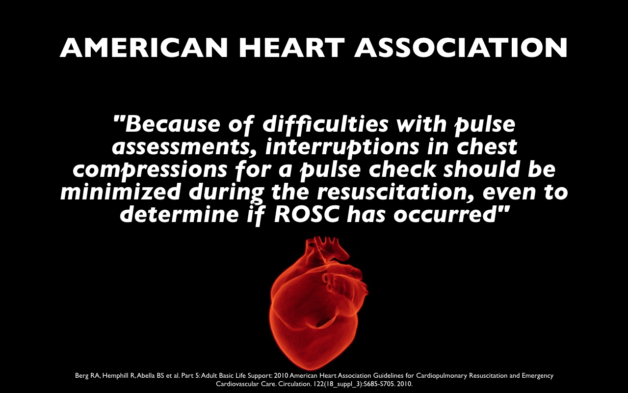
"Because of difficulties with pulse assessments, interruptions in chest compressions for a pulse check should be minimized during the resuscitation, even to determine if ROSC has occurred" [4].


It's really difficult to identify pulselessness in < 10 seconds
. Few people can determine the presence of pulselessness in 10 seconds. Dick et al performed a study of patients placed on cardiopulmonary bypass, and providers were blinded to the presence of pulsatile flow. Only two percent of experienced providers (n=209) were able to determine that a patient was pulseless in under 10 seconds [5].
Enforcement of time between compressions may be mitigated by having someone count down from 10 during the rhythm analysis. We often have the individual performing chest compressions do this and they are trained and reminded at the beginning of the resuscitation to resume compressions when they reach zero.

Pulse checks are inaccurate. A study by Tibaballs et al again had providers assess for a pulse in patients on bypass with and without pulsatile flow. They found 78% accuracy in identification of the presence or absence of a pulse [6]. While an accuracy of 78% may seem high, this means that approximately one in four times we are wrong. This means we may feel the reverberation of our own pulse and the truly pulseless patient may have an unnecessary and perhaps deleterious delay in chest compressions. Cardiac ultrasound and arterial line tracings demonstrate contractility and flow with superior diagnostic characteristics, although each has their limitations [7].

Why would we undertake a diagnostic strategy if we know it it is inaccurate and insensitive? Changing practice is difficult. In critical situations, we default to what is familiar, what we know.  It is time to move away from the pulse check. We may still need to check for rhythm analysis at periodic intervals, and this is supported by the AHA guidelines. Some monitoring systems allow for this concurrently with compressions (filter out the baseline), while others do not. Many of us are also compelled to search for reversible causes of arrest with ultrasound, many of which do not require an interruption in compressions.

How do we determine ROSC, then?
The AHA recommends arterial lines, ultrasound, rise in end-tidal capnography to 35-45 mmHg, or pulse AND blood pressure. In the emergency department, we often have access to ultrasound, arterial lines, and ETCO2.

If you're curious about why we are so stubborn to let go of our practices, check out this post on unlearning practices we adore.

References:
1. Soar J et al. "European Resuscitation Council Guidelines for Resuscitation 2015: Section 3. Adult advanced life support." Resuscitation, October 2015, Pages 100 - 1472.
2. Eftestøl T, Sunde K, Steen PA.  Effects of Interrupting Precordial Compressions on the Calculated Probability of Defibrillation Success During Out-of-Hospital Cardiac Arrest. Circulation.2002; 105: 2270-2273.
3. Link MS, Berkow LC, Kudenchuk PJ et al. Part 7: Adult Advanced Cardiovascular Life Support. Circulation. 132(18 suppl 2):S444-S464. 2015
4.Berg RA, Hemphill R, Abella BS et al. Part 5: Adult Basic Life Support: 2010 American Heart Association Guidelines for Cardiopulmonary Resuscitation and Emergency Cardiovascular Care. Circulation. 122(18_suppl_3):S685-S705. 2010.
5. Dick WF, Eberle B, Wisser G, Schneider T. The carotid pulse check revisited: what if there is no pulse? Crit Care Med. 2000 Nov;28(11 Suppl):N183-
6. Tibballs J, Weeranatna C. The influence of time on the accuracy of healthcare personnel to diagnose paediatric cardiac arrest by pulse palpation. Resuscitation. 81(6):671-5. 2010.
7. Gaspari R et al. Emergency department point-of-care ultrasound in out-of-hospital and in-ED cardiac arrest. Resuscitation 2016 Sep 27

Unlearning - Yoga For Your Brain


The Gist:  Knowledge translation is a problem in medicine and, at the individual level, unlearning likely contributes to the knowledge translation gap.  It may also exist as part of the solution. Akin to yoga, unlearning requires flexibility, training or deliberate practice, and is enhanced by a community open to skepticism and growth.
  • Note: These musings are not evidence based but are more of a cognitive framework to understand why we have such difficulty individually changing practice.
For an introduction to unlearning, check out this post.  In brief, unlearning, while really an aspect of truly understanding and learning, is complicated.  Most of us find it relatively easy to stuff more information into our brains, a process we perceive as "learning."  Nasal cannula with oxygen at 15 liters per minute plus may help prolong safe apnea time and reduce hypoxia during intubation?  Cool, we can do that.  However, when we are told that something that we routinely do or believe may not necessary or even harmful, we often have more difficulty changing out behavior or "unlearning." Intubating patients with out of hospital cardiac arrest may not be helpful? Preposterous! Unlearning a bit of information is unnatural, it may feel awkward whereas learning, as it often does not put our knowledge or ego in jeopardy.

Flexibility - Unlearning requires cognitive flexibility. When we stretch our bodies, we reach a point where we feel a burn; our body telling us we are approaching our limit. We can push further safely, it just burns slightly more.  Unlearning is essentially the same and is often accompanied by “the burn.”  When we come across information counter to the way we practice, it stings. We may feel defensive. This is “the burn.”  We experience discomfort when we stretch our thinking beyond this. This may be, in part, because we are emotionally tied to our knowledge. We work hard for what we know.  We act quickly in emergency care and must have confidence in what we know, as emergent situations do not typically allow for debate or significant time to think.

  • A fix: When one feels “the burn” when confronted with new information that runs counter to our practice, recognize that this is a warning sign that knowledge may be changing.  Recognize that the sting comes from our ego, protecting what we know.  This does not mean that one should change practice whenever we come across a piece of novel information. Rather, we should we aware that in order to practice evidence based, up to date medicine, we may feel discomfort. When our beliefs are challenged, instead of becoming defensive, we should thoughtfully consider the information.
Training - Mastering a yoga pose requires training and deliberate practice. In order to unlearn ways of thinking, we must also engage in mental preparation and practice.  It is easy, particularly in emergency settings when adrenaline dominates, to think and execute in a perfunctory manner. We default to what we know and what is familiar.
Some fixes:

  • Early Exposure - The earlier we begin training, the more prepared we are. If one has an upcoming race, we may expect we will perform better if we begin preparing earlier rather than the week before the race. Similarly, when it comes to unlearning a habit or a way of thinking, the sooner we are exposed to the contrary argument, the more prepared we may be to unlearn.  This may serve as a preconditioning so that we may react less strongly upon repeat exposure. 
  • Repeat Exposure - Practice is central to most athletic endeavors. The more repetitions we do, the stronger we become.  The more we practice a yoga pose, the more likely we are to be successful, the more comfortable it will feel.  Unlearning is easier when we are exposed to the target bit of knowledge more frequently.  Spaced repetition exists as one of the most evidence-based means of learning and  this probably applies to unlearning. 
Community - Yoga and CrossFit are associated with strong communities, as are many team sports.  Communities may motivate us, hold us accountable, and push the bounds of our perceived capability.  Studies demonstrate that physicians practice similarly to the institution where they trained and show a wide array of geographic variation in practice patterns. A network of peers and colleagues, particularly outside of own’s main “system” or hospital, may increase our cognitive flexibility by exposing us to a wide array of practice pattern.  The Free Open Access Medical Education (FOAM) community may expose to novel and controversial information.

Unlearning in the Prehospital Arena: The Workout
Needle Decompression for Tension Pneumothorax (see this post or this podcast).  The second intercostal space at the midclavicular line (2nd ICS MCL) has been taught as the ideal spot for needle decompression.  This, however, is changing.  New recommendations are to use a catheter at least 8cm in length if needle decompression is attempted at the 2nd ICS MCL or decompress at the fourth or fifth intercostal space at the anterior axillary line (4/5th ICS AAL).
The chest wall is thick at the 2nd ICS MCL [1,2].  Radiographic studies of chest wall thickness demonstrate increased thickness at the 2nd ICS MCL compared with the 4/5th ICS AAL (4.78 cm vs 3.42 cm).  Even ATLS states that needle decompression in the 2nd ICS MCL will fail more than 50 percent of the time.  This is an intervention undertaken in extreme circumstances in critically ill patients.  A chance of failure of 1 in 2 is unacceptable.
The 2nd ICS MCL is difficult to identify [3,4].  The clavicle extends further than most people think. As a result, providers are less accurate in identifying the 2nd ICS MCL compared with the 4/5th ICS AAL.


The pulse check.  If one were to survey cardiac arrest resuscitation across the United States, in and out of hospital,  we would probably see that the majority of people pause every two minutes for a “pulse check” despite decreased emphasis on the pulse check by the AHA guidelines over the past 10 years.  The guidelines recommend minimal interruptions for pulse check and detail the problematic sensitivity and specificity of pulse identification [6].  After the initial pulse check prior to CPR, the guidelines don’t actually specify any time frame for repeat pulse checks. Yet, many of us do.  Sure, we can pause for rhythm analysis; however, many systems and the European guidelines now recommend pulse assessment upon observation of an organized rhythm or increase in end tidal capnography [7].
Few people can determine the presence of pulselessness in 10 seconds. Dick et al of patients placed on cardiopulmonary bypass and providers blinded to whether or not the patient actually had a pulse. Only 2% of this cohort of experienced providers were able to identify a pulseless patient in 10 seconds [8]. With increased emphasis on compression fraction, this may result in a delay in resumption of compressions.
The accuracy of the pulse identification by providers is suboptimal, noted to be 78% in one study [9].  While an accuracy of 78% may seem high, this means that approximately one in four times we will be wrong. We may feel the reverberation of our own pulse and the truly pulseless patient may have an unnecessary and perhaps deleterious delay in chest compressions.
For more on this topic check out this post and/or this post.


Left Bundle Branch Block (LBBB) as a STEMI Equivalent (check out this post)- Prior to the 2013 iteration of the AHA guidelines for ST-elevation myocardial infarction (STEMI), new or presumed new LBBB existed as a “STEMI equivalent.”  This often activated the cath lab and STEMI teams.  In 2013, the AHA removed this from the guidelines yet these patients are often referred to the emergency department for “rule out MI.”
Further, STEMI may often be diagnosed on ECG, using the Sgarbossa or modified Sgarbossa criteria (link) [10].


Backboards - fortunately protocols in many states and systems have dispensed with long backboards.  Long thought to be protective, despite known harms, the American College of Emergency Physicians released a guideline in 2016 explicitly stating that long backboards should not be used as a therapeutic or precautionary measure. They cause harm and don’t help. [11]

Oxygen in Acute Coronary Syndromes - Aspirin, oxygen, and nitroglycerin have long been the initial interventions for patients with suspected ACS. Recent studies have found no clear benefit for oxygen in patients with normal oxygen saturations. Further, one study found oxygen was associated with markers of larger myocardial infarctions (although this is not a patient-oriented outcome) [12]. The AHA recommends oxygen is appropriate for patients who are hypoxemic (oxygen saturation < 90%) [6].


References:
1.  Laan D V., Vu TDN, Thiels CA, et al. Chest wall thickness and decompression failure: A systematic review and meta-analysis comparing anatomic locations in needle thoracostomy. Injury. 2015:14–16.
2. Advanced Trauma Life Support, 9th edition.
3. Ferrie EP et al. The right place in the right space? Awareness of site for needle thoracocentesis. Emerg Med J 2005;22(11):788–9.
4. Inaba K et al. Cadaveric comparison of the optimal site for needle decompression of tension pneumothorax by prehospital care providers
5. ACS COT
6. Berg RA, Hemphill R, Abella BS et al. Part 5: Adult Basic Life Support: 2010 American Heart Association Guidelines for Cardiopulmonary Resuscitation and Emergency Cardiovascular Care. Circulation. 122(18_suppl_3):S685-S705. 2010.
7. Resuscitation. 81(6):671-5. 2010.
8. Dick WF, Eberle B, Wisser G, Schneider T. The carotid pulse check revisited: what if there is no pulse?Crit Care Med. 2000 Nov;28(11 Suppl):N183-5
9. Tibballs J, Weeranatna C. The influence of time on the accuracy of healthcare personnel to diagnose paediatric cardiac arrest by pulse palpation.
10. O'Gara PT, Kushner FG, Ascheim DD et al. 2013 ACCF/AHA Guideline for the Management of ST-Elevation Myocardial Infarction. Journal of the American College of Cardiology. 61(4):e78-e140. 2013
11. "EMS Management of Patients with Potential Spinal Injury." ACEP Board of Directors. Available at: https://www.acep.org/clinical---practice-management/ems-management-of-patients-with-potential-spinal-injury/
12. Stub D et al. Air Versus Oxygen in ST-Segment-Elevation Myocardial Infarction. Circulation. 131(24):2143-2150. 2015.

Saturday, May 21, 2016

P Values: Everything You Know Is Wrong

The Gist:  P values are probably the most “understood” statistic amongst clinicians yet are widely misunderstood.  P values should not be used alone to accept or reject something as “truth” but they may be thought of as representing the strength of evidence against the null hypothesis [1].

Part of our 15 Minutes - 'Stats are the Dark Force?' residency lecture series

P: Everything You Know Is Wrong from Lauren Westafer on Vimeo.


At various times in my life I, like many others, have believed the p value to represent one of the following (none of which are true):
    • Significance
      • Problem:  Significance is a loaded term.  A value of 0.05 has become synonymous with “statistical significance.”  Yet, this value is not magical and was chosen predominantly for convenience [3].  Further, the term “significant” may be confused with clinical importance, something a statistic cannot answer.
    • The probability that the null hypothesis is true.
      • Problem: The calculation of the p value includes the assumption that the null hypothesis is true.  Thus, a calculation that assumes the null hypothesis is true cannot, in fact, tell you that the null hypothesis is false.
    • The probability of getting a Type I Error 
      • Background: Type I Error is the incorrect rejection of a true null hypothesis (i.e. a false positive) and the probability of getting a Type I Error is represented by alpha. Alpha is often set at 0.05 so that there is a 5% chance you are wrong if you reject the null hypothesis. This is a PRE test calculation (set before the experiment)
      • Problem: Again, the calculation of the p value assumes the null hypothesis is true. The p value only tells us the probability of getting the data we did, it does NOT speak to the underlying truth of whatever is being tested (i.e. efficacy). The p value is also a POST test calculation.
      • The error rate associated with various p values varies, depending on the assumptions in the calculations, particularly prevalence. However, it's interesting to look at some of the estimates of false positive error rates often associated with various p values:  p=0.05 - false positive error rate of 23-50%; p=0.01 - false positive error rate of 7-15% [5].
P value is the probability of getting results as extreme or more extreme, assuming the null hypothesis is true. Originally, this statistic was intended to serve as a gauge for researchers to decided whether or not a study was worth investigating further [3].  
  • High P value - data are likely with a true null hypothesis [Weak evidence against the null hypothesis]
  • Low P value - data are UNlikely with a true null hypothesis [Stronger evidence against the null hypothesis]
Example:  A group is interested in evaluating needle decompression of tension pneumothorax and proposes the following:
    • Hypothesis - Longer angiocatheters are more effective than shorter catheters in decompression of tension pneumothorax.
    • Null hypothesis - There is no difference in effective decompression of tension pneumothorax using longer or shorter angiocatheters.
A group, Aho and colleagues, did this study and found a p value of 0.01 with 8 cm catheters compared with 5 cm catheters.  How do we interpret this p value?  
  • We would expect the same number of effective decompressions or more in 1% of cases due to random sampling error.  
  • The data are UNLIKELY with a true null hypothesis and this is decent strength evidence against the null hypothesis.
Limitations of the Letter “P”
    • Reliability.  P values depend on the statistical power of a study. A small study with little statistical power may have a p value greater than 0.05 and a large study may reveal that a trivial effect has statistical significance [2,4].  Thus, even if we are testing the same question, the p value may be "significant" or "nonsignificant" depending on the sample size.
    • P-hacking.  Definition:  "Exploiting –perhaps unconsciously - researcher degrees of freedom until p<.05" Alternatively: "Manipulation of statistics such that the desired outcome assumes "statistical significance", usually for the benefit of the study's sponsors" [7].
      • A recent study of abstracts between 1990-2015 showed 96% contained at least 1 p value < 0.05.  Are we that wildly successful in research? Or, are statistically nonsignificant results published less frequently (probably).  Or, do we try to find something in the data to report as significant, i.e. p-hack (likely).
P values are neither good nor bad. They serve a role that we have distorted and, according to the American Statistical Association: The widespread use of “statistical significance” (generally interpreted as “p ≤ 0.05”) as a license for making a claim of a scientific finding (or implied truth) leads to considerable distortion of the scientific process [1].  In sum, acknowledge what the p value is and is not and, by all means, do not p alone.

References:
  1. Wasserstein RL, Lazar NA. The ASA’s statement on p-values: context, process, and purpose. Am Stat. 2016;1305(April):00–00. doi:10.1080/00031305.2016.1154108.
  2. Goodman S. (2008) A dirty dozen: twelve p-value misconceptions. Seminars in hematology, 45(3), 135-40. PMID: 18582619 
  3. Fisher RA. Statistical Methods for Research Workers. Edinburgh, United Kingdom: Oliver&Boyd; 1925.
  4. Sainani KL. Putting P values in perspective. PM R. 2009;1(9):873–7. doi:10.1016/j.pmrj.2009.07.003.
  5. Sellke T, Bayarri MJ, Berger JO.  Calibration of p Values for Testing Precise Null Hypotheses.  The American Statistician, February 2001, Vol. 55, No. 1
  6. Chavalarias D, Wallach JD, Li AHT, Ioannidis JPA. Evolution of Reporting P Values in the Biomedical Literature, 1990-2015. Jama. 2016;315(11):1141. doi:10.1001/jama.2016.1952.
  7. PProf.  "P-Hacking."  Urban Dictionary. Accessed May 1, 2016.  Available at: http://www.urbandictionary.com/define.php?term=p-hacking

Tuesday, January 26, 2016

Percutaneous Chest Tubes: The Humane Choice

The Gist:  Small bore percutaneous catheters, often referred to as "pigtail" catheters, should be the initial means of treating many pneumothoraces and select other drainable thoracic pathologies as they cause less pain and capitalize on the commonly used seldinger technique [1-10].

Traditional tube thoracostomy is an invasive procedure.  For the past several years, international guidelines, individuals in the Free Open Access Medical education (FOAM) community, and various institutions have moved towards placing more pigtail catheters for urgent thoracic pathology.  Yet, this practice is still not ubiquitous.  I recently gave a talk on this to my program and, in the spirit of FOAM, have shared it:

Technique - Watch this video by Dr. Larry Mellick 
  • Seldinger style: uses a technique with which we are intimately familiar. The majority of emergency providers have likely done far more central lines than open tube thoracostomies. As such, a technique mentally and mechanically familiar to providers may be preferable.
  • Pearls for placement - Kulvatunyou and colleagues suggest "POW" pearls for placement.
    • P -Perpendicular: Ensure the finder needle is perpendicular to the rib during placement
    • O -Over the rib: Like chest tubes, pigtails go over the rib to avoid injury to the neurovascular bundle
    • W -Wary of wire kinking:  The wire may be prone to kinking, particularly upon dilation through the tough intercostal muscles.
Indications
  • Pneumothorax
    • Spontaneous pneumothorax: The British Thoracic Society has recommended small bore tubes over traditional chest tubes since 2010.
    • Traumatic pneumothorax:  Use of pigtail catheters have increased in many trauma communities, with success rates comparable to large bore chest tubes [8-11].
  • Effusions - pigtail catheters are frequently used to drain effusions, particularly simple effusions. Most of the primary literature on this topic has been conducted in children with parapneumonic effusions and has demonstrated that this technique is successful and safe [13].
Cautions/Contraindications
  • Hemothorax/Complex fluid - Larger bore tubes (28F and larger) are typically used to drain hemothorax due to the feared complication of retained hemothorax.  A prospective review of 36, 14F pigtail catheters placed for hemothorax in trauma patients found no significant differences in complications or success between pigtails or chest tubes but wasn't powered to find important, infrequent complications [11].  An animal study found
The Good:
  • Less Painful - In addition to the procedure not requiring large, forceful separation of the and unsurprisingly, placing a smaller tube in the chest causes less pain, even 2 days after the procedure [10].
    • Pain in pigtail vs chest tube patients: Day 0 3.2  vs 7.7; p<0.001, Day 1 1.9 vs 6.2; p<0.001, Day 2 2.1 vs 5.5; p=0.04 (note: no power calculation performed)
  • Easy/Familiar Procedure - as above under "Seldinger technique"
  • May reduces some complications - The literature suggests that complications are typically at least equivalent between larger chest tubes and pigtails. More serious complications are difficult to quantify given the infrequency.
    • One study did show that infections were reduced in the pigtail group, possibly due to technique or a larger nidus for infection [2].
  • Outpatient treatment possible - In select patient groups with spontaneous pneumothorax and excellent follow up, a pigtail catheter may be connected to a heimlich valve and the patient may be discharged [7].
The Bad:
  • More predisposed to kinking - Due to the small, flexible tubing, these tubes may kink and obstruct the lumen.  The trauma literature suggests these complications may occur in 2-8% of cases [8-10].
  • Clogging - Drainage of some complex fluids (loculated effusion/hemothorax) may be more problematic through pigtail catheters as the small lumen may be easier obstructed with clot.
  • Time? The belief exists that open thoracostomy more expediently relieves pneumothorax compared with the percutaneous technique and is thus preferred in emergent, life-threatening situations. To date, there's no literature to support or refute this and the time a tube takes is likely provider dependent.
  • It's less cool - A certain pride and thrill exists with performing invasive procedures.  In discussions with individuals regarding barriers to uptake of the percutaneous technique the theme arose that performing this technique would demonstrate some sort of weakness by the provider. Note: this notion is not supported or addressed by the literature and is merely a thought about subconscious provider bias
References:
1. Laws D et al. BTS guidelines for the insertion of a chest drain. Thorax. 2003 May;58 Suppl 2:ii53-9.
2.  Benton IJ, Benfield GF. Comparison of a large and small-calibre tube drain for managing spontaneous pneumothoraces. Respir Med. 2009 Oct;103(10):1436-40.
3. Dull KE, Fleisher GR. Pigtail catheters versus large-bore chest tubes for pneumothoraces in children treated in the emergency department. Pediatr Emerg Care. 2002 Aug;18(4):265-7.
4. Gammie JS et al. The pigtail catheters for pleural drainages: a less invasive alternative to tube thoracostomy. JSLS. 1999 Jan-Mar;3(1):57–61.
5. Kuo HC, et al. Small-bore pigtail catheters for the treatment of primary spontaneous pneumothorax in young adolescents. Emerg Med J. 2013 Mar;30(3):e17.
6.  Repanshek ZD, Ufberg JW, Vilke GM, Chan TC, Harrigan RA. Alternative Treatments of Pneumothorax. J Emerg Med. 2013 Feb;44(2):457-466.
7. Hassani B, Foote J, Borgundvaag B. Outpatient management of primary spontaneous pneumothorax in the emergency department of a community hospital using a small-bore catheter and a Heimlich valve. Acad Emerg Med. 2009 Jun;16(6):513-8.
8. Kulvatunyou N, Vijayasekaran A, Hansen A, et al. Two-year experience of using pigtail catheters to treat traumatic pneumothorax: a changing trend. J Trauma. 2011 Nov;71(5):1104-7.
9. Rivera L, O’Reilly EB, Sise MJ, et al. Small catheter tube thoracostomy: effective in managing chest trauma in stable patients. J Trauma. 2009 Feb;66(2):393–9
10.  Kulvatunyou N, et al. A prospective randomized study of 14-French pigtail catheters vs 28F chest tubes in patients with traumatic pneumothorax: impact on tube-site pain and failure rate. EAST Annual Surgical Assembly, Oral paper 12, Jan 17, 2013.
11. Kulvatunyou N, Joseph B, Friese RS, et al. 14 French pigtail catheters placed by surgeons to drain blood on trauma patients. J Trauma Acute Care Surg. 2012;73(6):1423–1427. 
12. Russo RM, Zakaluzny SA, Neff LP, et al. A pilot study of chest tube versus pigtail catheter drainage of acute hemothorax in swine. J Trauma Acute Care Surg. 2015;79(6):1038–1043. 
13.  Liu YH, et al. Ultrasound-guided pigtail catheters for drainage of various pleural diseases. Am J Emerg Med. 2010 Oct;28(8):915-21
14. Inaba K, Lustenberger T, Recinos G. Does size matter? A prospective analysis of 28-32 versus 36-40 French chest tube size in trauma. The journal of trauma and acute care surgery. 72(2):422-7. 2012.

Monday, December 29, 2014

Medicine's Third: Polypharmacy

The Gist: Polypharmacy, the concurrent use of multiple medications (5+) or use of unnecessary medications, is problematic in medicine.  Consider “medication related problem” on the differential diagnosis and review the patient’s medications.  When prescribing a medication, consider the unwanted reactions and tailor therapy, recalling that medications frequently have subtle or additive effects that may be especially problematic in the elderly. When in doubt, send a communication to a patient's PCP.

The Case: A 58 y/o with a history of hypertension and diabetes presented with weakness, vomiting, and fatigue.  A basic chemistry panel returned with a creatinine of 3.8 mg/dL (last value, 0.9 mg/dL).  While initially it seemed as though the gentleman had prerenal acute kidney injury from vomiting, the patient revealed he had been taking both ibuprofen and naproxen for worsening arthritis, in addition to his prescribed ace-inhibitor and thiazide diuretic.  See another case in this post on medication reconciliation.

Newton's Third Law states:
"For every action there is an equal and opposite reaction.” 

We ponder this frequently looking at collisions or calculating billiard shots but I think this principle can be translated to medicine. In the medical realm we prescribe therapies for the primary action of that medication/intervention.  Yet, unintended consequences abound.  Despite the comically long “disclaimers” of side effects on advertisements, the additive effects, unintended as they may be, are often disguised in a patient’s presenting complaint.  Further, patients are often prescribed medication to mask the side effects of another medication. Struck by this during medical school, I created my own version:
Westafer’s Third Law of Medicine:
 “For every medication action there is an unequal and unintended reaction.” 

This came up recently in a discussion on Twitter regarding a new medication for hyperkalemia, targeted to combat the side effect of elevated potassium in patients on ACE-inhibitors, ZS-9. A medication for a medication side effect (with likely more broad application in reality).


Although prescriptions from the emergency department (ED) are likely a minority of offenders with regard to the volume of inappropriate medications, awareness of the role that medications may play in the patient’s complaint. Studies show that adverse drug events (ADEs) may be responsible for 10-12% of ED visits among patients > 65 years old, although the definition of adverse drug event and determination of causality vary based on the study [1-3].   A more recent Canadian database review demonstrated a lower prevalence of ADEs generating ED visits, 0.8%, but the methods leave something to be desired [4].

A small study by Chin and colleagues identified ED prescriptions for analgesia, notably NSAIDs, muscle relaxants, and narcotics, as an area for future intervention [5].  Interestingly, this paper was published prior to the massive spike in opioid prescriptions; thus, this area may be even more crucial presently [6].  

Deleterious
Polypharmacy, particularly in the elderly, is associated with an increase in the prevalence of falls, mortality, hospital admission, and hospital length of stay.  The elderly are more susceptible to many of these effects as clearance and metabolism change with age, and elderly patients tend to be on more medications. 

Drug-drug interaction - A medication alters the activity of another.  Example: warfarin + ciprofloxacin -> supratherapeutic INR and may lead to increased bleeding.
Drug-disease interaction - Medications that should be avoided in patients with specific medical conditions.  
  • Example: Use of aspirin 325 mg or non-steroid anti-inflammatories in patients with peptic ulcer disease.
Adverse effects - Many medications have more pronounced adverse effects in elderly patients, often because the pharmacokinetics, such as renal excretion, are altered and may predispose patients to acute kidney injury, delirium, or orthostatic hypotension.  Check out this podcast for more.
  • Example: Anticholinergic properties are abundant in medications, including antidepressants, antihistamines, and antipsychotics.  In the elderly these effects are more pronounced and are associated with hallucinations, impaired memory, tachycardia, falls, constipation, etc.
Unnecessary - Medications are frequently initiated and then continued without re-examination for appropriateness. A study of Veterans Association hospital discharges of patients age >65 y/o classified as "frail" found that 44% had at least one unnecessary medication at discharge [8]. These medications contribute to increase cost and may play a role in further drug interactions or adverse effects. 
  • Example: A H2 blocker such as ranitidine may be prescribed for prophylaxis but the anticholinergic effects can contribute to diminished cognition, constipation, etc (see above).
Under-recognized  A prospective observational study by Hohl and colleagues of ED patients > 65 y/o in Canada found ADEs in 8.3%-12.3%, depending on the breadth of the definition of ADEs.  A prospective study by Hohl et al found that many ADEs in the ED were not attributed as medication related, particularly in the older population [9]. 

ED Interventions
  • Consider the Third Law of Medication when pondering the differential diagnosis.  For example, geriatric fall patients should probably be screened for polypharmacy (What medications is the patient on?  Can the problem be explained by a medication?) and while prescribing medications (Is the medication truly necessary? Will it interact with any of their medications?  Does the patient need a bowel regimen or other precautions?)
  • Medication review in the ED.  The ED encounter can serve as an opportunity for an outsider to glance at the patients medications to gain a sense as to whether something may be dangerous or warrant further discussion with their primary physician.
  • Judicious prescription of medications.  In the ED, we often write for short courses of medication and may be lulled into the sense that these prescriptions don't matter, yet they may carry an unintended reaction.  Be familiar with medications that are common offenders.
    • The Beers' List has a long list of medications to avoid in the elderly, but often these aren't the biggest offenders (also note the STOPP criteria). The most common medications associated with ADEs, implicated in 67% of hospital admissions according to a national survey database, were: 
      • warfarin (33.3%)
      • insulins(13.9%)
      • oral antiplatelet agents (13.3%)
      • oral hypoglycemic agents (10.7) [1,4]
  • Targeted feedback to general practitioners regarding potentially problematic medications.  Many health systems and electronic medical records have easy ways to send messages to primary care physicians.  In the ED haste, these communications frequently take a back seat but may be important.  Yet, the ACEP Geriatric ED guidelines recommend referral to PCP for any concern for polypharmacy (>5 medications) or presence of any high risk medication [10].
  • ED pharmacists. Many study authors have called for increasing the role of ED pharmacists in identifying ADE related to medications [2].
References:
1.Budnitz DS, Lovegrove MC, Shehab N, Richards CL. Emergency hospitalizations for adverse drug events in older Americans. N Engl J Med. 2011;365:(21)2002-12. 
2. Banerjee A, Mbamalu D, Ebrahimi S, Khan AA, Chan TF. The prevalence of polypharmacy in elderly attenders to an emergency department - a problem with a need for an effective solution. Int J Emerg Med. 2011;4(1):22.
3. Budnitz DS, Shehab N, Kegler SR, Richards CL. Medication use leading to emergency department visits for adverse drug events in older adults. Ann Intern Med. 2007;147:(11)755-65.
4.Bayoumi I, Dolovich L, Hutchison B, Holbrook A. Medication-related emergency department visits and hospitalizations among older adults. Can Fam Physician. 2014;60:(4)e217-22. 
5. Chin MH, Wang LC, Jin L, et al. Appropriateness of Medication Selection for Older Persons in an Urban Academic Emergency Department. Acad Emerg Med. 2007;6(12):1232–1242.
6. Ruscitto A, Smith BH, Guthrie B. Changes in opioid and other analgesic use 1995-2010: Repeated cross-sectional analysis of dispensed prescribing for a large geographical population in Scotland.Eur J Pain. 2015 Jan;19(1):59-66. 
7. Robitaille C, Lord V, Dankoff J, et al. Emergency Physician Recognition of Adverse Drug-related Events in Elder Patients Presenting to an Emergency Department. 2005;12(3). 
8.Hajjar ER, Hanlon JT, Sloane RJ, et al. Unnecessary drug use in frail older people at hospital discharge. J Am Geriatr Soc. 2005;53:(9)1518-23. 
9. Hohl CM, Zed PJ, Brubacher JR, Loewen PS, Purssell RA. Do Emergency Physicians Attribute Drug-Related Emergency Department Visits to Medication-Related Problems? YMEM. 2009;55(6):493–502.e4. 
10.American College of Emergency Physicians. Geriatric emergency department guidelines 2013

Saturday, November 22, 2014

Misrepresented: EBM

The Gist: Evidence based medicine (EBM) is misunderstood; it's not a randomized control trial (RCT) or "the literature." Rather, EBM is the intersection of the best available evidence, clinical expertise, and patient values [1-2]. Avoid BARF (Brainless Application of Research Findings), with tips from Emergency Medicine Cases

We have a cultural problem.  Clinicians are increasingly called upon to practice EBM.  Yet, the term EBM does not sit well on the palate of many physicians.  Conversations involving a mention of EBM have resulted in some of the following refrains...
"See, my patients are different..." 
"We'll never get an RCT on that..." 
"The culture is different here, I don't want to get sued." 
"Patients don't understand, but they do hold the power with satisfaction scores." 
"It's cookbook medicine."
With these words and reactionary body language, the dialogue quickly shuts down - by both parties.  First, this is a shame.  We should learn from one another but there seems to be a "hard stop" between many who champion EBM and those who find EBM off-putting. Second, this is a misunderstanding.  EBM is not an RCT.  In fact, EBM is not the best statistical methods or the rationing of care. EBM is not nihilism.  

EBM is the intersection of the best available evidence, clinical expertise, and patient values:
"the conscientious, explicit, and judicious use of current best evidence in making decisions about the care of individual patients. The practice of evidence based medicine means integrating individual clinical expertise with the best available external clinical evidence from systematic research" [1].
Why, then, the misunderstanding? 
Here are some thoughts...

Misrepresentation. EBM is often used to refer to literature or studies, rather than to the application of research and evidence to particular patients and situations, using one's clinical experience (example and discussion: "EBM is Crap").  As a result, EBM may be misunderstood as a cost-cutting venture or a cookbook for medicine [3]. I have been complicit in perpetuating this misrepresentation of EBM.   As a novice physician-in-training with limited clinical experience, I draw predominantly upon the literature base.  I have unknowingly quoted the literature, thereby proudly proclaiming my practice of EBM, while unconsciously dismissing the other components of EBM.  
  • A remedy:  Remind ourselves and others that the evidence is part of the trifecta of EBM, along with the patient's values and clinical expertise.  We can be clear in what we mean by EBM and refrain from referring to a body of literature as EBM. 
Zeal. A religiosity exists amongst many champions of EBM, or people who believe they are championing EBM.  We tout our pyramids of evidence and may scoff at a lack of evidence or rigorous trials.  This may be off-putting as not all fields are amenable to RCTs and patient populations vary.  Moreover, there's a human tendency to form a reactionary attitude when someone exerts a strong identity [4].  Hence, EBM zeal may engender an anti-EBM attitude and cause people to be wary of solid practice changing evidence.
  • A remedy:  While championing good research and employing the best available evidence, we can balance our enthusiasm with important caveats and understand the importance for tailored approaches for patients.  Gentle education about EBM rather than diatribes may aid individuals in understanding the values of EBM beyond evidence.
Fear.  People like to be right.  We may reflexively become defensive when we are (possibly) wrong. EBM or "literature" can be used in an antagonizing way and, subconsciously, a way to exert a feeling of superiority.  "You haven't read that study?"
  • A remedy: Understand that unlearning in medicine is difficult.  Aggressive assertions may push people further away.  Think of it as a Kubler-Ross like grief cycle, as explained in this post.  This may help us become more cognitively flexible, understand the reticence of others, and perhaps make our points more effectively.  


Confusion.  Historically, researchers, clinicians, physicians in training, and allied health professionals have limited understanding of fundamental statistics [5,6].  As such, we may not understand what we're reading or how it applies to our patient population.  We may have difficulty understanding why something we believed was proper at one time is no longer the case.  Often, this is because the research was, in fact, initially wrong or misleading [7]. 
  • A remedy: Read.  This podcast proffers tips on getting started; however, even the most seemingly rigorous papers published in high impact journals are subject to bias (publication bias and otherwise), which can be difficult to parse through.  For example, the oseltamivir (tamiflu) recommendations from Cochrane changed after they were allotted access to data, demonstrating the profound impact of publication bias [Jefferson et al].  More on this here.
Time. The number of journal articles needed to read (NNR) to obtain valid and relevant information is typically cited as 20-200, an insurmountable task [8].  The process of trolling through the literature is time consuming and may be overwhelming.  Frustration can turn into apathy, confusion, and mistrust.
There are legitimate issues with EBM.  Evidence is often subject to the biases of industry and legislative bodies.  Guidelines or recommendations billed as "EBM" may be hijacked by individuals with conflicts of interest or other agendas. Further, the grading of evidence isn't always objective or consistent, as seen by the grading of evidence for thromboylitics in acute ischemic stroke listed in the ACEP clinical policy.   In addition, guidelines harness EBM and disseminate the body of evidence to practitioners.  For example, the 2008 AHA/ACC guidelines are based largely on low levels of evidence and expert opinion,  many of whom have financial conflicts of interest.  Only 11% of the recommendations were based on high quality evidence [9].  

So, while EBM has imperfections in concept, representation, and implementation, the model incorporates the primary things we, as providers, care about - the evidence, the patient, and clinical experience.  Let's understand what EBM means and apply the term and principles appropriately.

References:
1. Sackett DL, Rosenberg WM, Gray JAM, et al. Evidence based medicine: what it is and what it isn’t. BMJ. 1996;312(7023):71–72. 
2. Greenhalgh T, Howick J, Maskrey N. Evidence based medicine: a movement in crisis? BMJ 2014;348:g3725
3. Straus SE, McAlister FA. Evidence-based medicine: a commentary on common criticisms. CMAJ. 2000;163(7):837–41. 
4.  Maalouf A.  In the Name of Identity: Violence and the Need to Belong. New York: Penguin Books, 2000.
5.  Windish D, Huot S, Green M. Medicine residents’ understanding of the biostatistics and results in the medical literature. Jama. 2007;298(9). 
6.  Mavros MN, Alexiou VG, Vardakas KZ, Falagas ME. Understanding of statistical terms routinely used in meta-analyses: an international survey among researchers. PLoS One. 2013;8(1):e47229. 
7.Ioannidis JP a. How many contemporary medical practices are worse than doing nothing or doing less? Mayo Clin Proc. 2013;88(8):779–81.
8. McKibbon KA, Wilczynski NL, Haynes RB. What do evidence-based secondary journals tell us about the publication of clinically important articles in primary care journals? BMC Med. 2004;2:33. 
9.  Tricoci P1, Allen JM, Kramer JM, et al.  Scientific evidence underlying the ACC/AHA clinical practice guidelinesJAMA. 2009 Feb 25;301(8):831-41.

Saturday, November 8, 2014

SBO Ultrasound

The GistAs mentioned in this post, the operating characteristics of historical and physical features are suboptimal in small bowel obstruction (SBO).  Bedside ultrasound has better operating characteristics and is one of the easier scans to perform and read.  Assuming others like to make their lives easier, I gave a talk on this; but professionals have created a tutorial at The Ultrasound Podcast tutorial.

I delivered a quick talk at the Controversies and Consensus in Emergency Medicine Conference on ultrasound for SBO, a modality that I've found great utility for in my developing practice. As a believer in Free Open Access Medical education (FOAM) and with hopes that, as a novice I might receive some constructive criticism to help me become better, I have posted the recording.


A Few Tidbits (some redundancy from prior post): 
Time.  Ultrasound for SBO is quick and easy and can be performed in conjunction with the history and physical exam in appropriate patients.  This may alleviate the time to definitive diagnosis (say CT or surgical evaluation), treatment, and/or disposition.*  Furthermore, sometimes we see things we don't expect on ultrasound.  Familiarity with US findings of SBO may make sense of dilated loops of bowel or altered peristalsis encountered during a gallbladder or aorta scan for abdominal pain.  Conversely, there are times when SBO may be suspected and a quick ultrasound may reveal an alternative diagnosis that may grossly change management (examples in talk).

X-rays are out for SBO.  Bedside ultrasound has better operating characteristics than plain films with fewer instances of equivocal results.  Sometimes plain films are crucial to evaluate for pneumoperitoneum but most patients with abdominal pain don't fall in this category.  Indeed, The American College of Radiology conclusion on plain films in suspected SBO
"In light of these inconsistent results, it is reasonable to expect that abdominal radiographs will not be definitive in many patients with a suspected SBO. It could prolong the evaluation period and add radiation exposure while often not obviating the need for additional examinations, particularly CT" [5].
Limitations.
  • Ileus vs. SBO - while US beats plain films with regard to percentage of ambiguous scans, ultrasounds can be equivocal as well.
  • Cause of obstruction/Transition point not well elucidated.  In patients with recurrent SBO from malignancy or adhesions and this may be less important to the managing team and surgeons often stop ordering CT scans if the presentation is consistent with prior presentations. 
  • Consultant access to images obtained at the bedside.
Note:  I have not included surgical consultants requiring a CT scan as part of the limitations.  The surgical literature recognizes the capacity of US to diagnose SBO, although this is not yet widely adopted [6].  However, despite common assumptions, surgeons don't require a CT scan for every recurrent SBO.  As a result, sometimes a positive ultrasound, followed by plain film, may be enough in these patients who will undergo conservative management.  Have a chat with each consultant, they're not always as inflexible as we make them out to be. 

*NCT02190981 pending with LOS as secondary outcome


References:
1.  Carpenter CR, Pines JM. The end of X-rays for suspected small bowel obstruction? Using evidence-based diagnostics to inform best practices in emergency medicine. Acad. Emerg. Med. 2013;20(6):618–20.
2.  Taylor MR, Lalani N. Adult small bowel obstruction. Acad. Emerg. Med. 2013;20(6):528–44.
3. Böhner H, Yang Q, Franke C, Verreet PR, Ohmann C. Simple data from history and physical examination help to exclude bowel obstruction and to avoid radiographic studies in patients with acute abdominal pain. Eur. J. Surg. 1998;164(10):777–84. 
4. Jang TB, Schindler D, Kaji AH.  Bedside ultrasonography for the detection of small bowel obstruction in the emergency department. Emerg Med J. 2011 Aug;28(8):676-8.
5. Katz DS, Baker ME, Rosen MP, Lalani T, et al, Expert Panel on Gastrointestinal Imaging. ACR Appropriateness Criteria® suspected small-bowel obstruction. Reston (VA): American College of Radiology (ACR); 2013. 10 p.
6.  
Maung AA, Johnson DC, Piper GL et al. Evaluation and Management of Small-Bowel Obstruction.  J Trauma. 73(5):S362-S369, November 2012

Saturday, October 11, 2014

Euboxia - Not Necessary (Or Necessarily Normal)

The Gist:  In medicine, we historically strive towards achieving values that fall within a reference range, or are normal, a phrase coined "euboxia" [1].  Targeting treatments to normalize values may not result in patient-oriented benefit and may cause harm.  We must also consider that normal values may not necessarily be normal for our patients.  Data fatigue, the exposure to copious data, may lead to ignoring values that are not flagged as abnormal, regardless of the appropriateness for a patient.
"'Euboxia' (from the Greek 'eu' meaning good, normal or happy, and 'box' from the tradition of writing physiological variables in boxes) is a colloquial word used in many North American and other hospitals to describe the state of apparent perfection aimed at by residents by the time they present their patients on morning rounds" - MC Meade [2].
Euboxia Is Not Always Necessary
Chris Nickson's Free Open Access Medical education (FOAM) post on Euboxia highlights some of the pitfalls with this obsession with normalcy. He also delivered a talk Euboxia and (ab)Normality at SMACC Gold which will hopefully be available on the SMACC podcast in the near future. A few examples include:
  • Hemoglobin transfusion trigger in anemia - Studies such as TRICC, CRIT, SOAP, and TRISS demonstrate that transfusion targets of more "normal" hemoglobin levels is not advantageous and may incur increased risks.  As such, transfusion triggers, in the absence of active myocardial ischemia, have moved to <7 g/dL while uptake of this trigger remains low in some communities [4]. 
  • Oxygen saturation in COPD - Unless patients are under duress, guidelines suggest patients with COPD have oxygen saturations targeted to 88-92% rather than the 98-100% more often associated with perfection [5]
  • Blood gas and saturations in ARDS - Guidelines for ventilation in patients with ARDS aim to protect the lungs using low tidal volumes and plateau pressures at the expense of allowing a pH of 7.20, permissive hypercapnia, and lower oxygen saturations of 88-95% (paO2 55-80 mmHg).  Correction of these lab abnormalities may come at the cost of additional lung damage by means of higher pressures or volumes and are thus discouraged [6].
Euboxia Is Not Necessarily Normal
Euboxia, however, may fool also practitioners into a false sense of security.  Failure to truly see a value that appears normal and isn't flagged, red, or outside of the box may be problematic. A few examples:

Normotension - Hypotension typically refers to systolic blood pressure <90 mmHg or a drop in systolic blood pressure >40 mmHg.  The latter part of this definition is often unable to be determined (due to lack of information) or forgotten.  The trauma literature seems to have solidified around the notion that the widely accepted definition of hypotension does not apply to many trauma patients, particularly those > 65 years old, and that 110 mmHg is probably a better cutoff [9-12].  While these recommendations have been out since 2011,  90 mmHg remains the common cut point for hypotension.
  • The CDC triage guidelines/"National Trauma Triage Protocol" have suggested <110 mmHg as the new hypotension guideline in patients > 65 years of age as multiple registry studies have demonstrated that an SBP <110 mmHg is associated with increased mortality and has an improved AUC compared with other blood pressure cut offs [9]. 
    • An abstract presented at AAST in 2014 found that patients >65 y/o with an SBP 90-109 mmHg had an odds of mortality of 9.7 (95% CI 8.7-10.8, p<0.01).  This survey study found improved, but terrible sensitivity for Trauma Center Need (ISS>15, ICU admit, urgent OR, or ED death) with the higher SBP cut-off [10].
Normal White Blood Cell Count (WBC) - Leukocytosis is often used as a predictor of infection/inflammation and historically loved by surgical services, yet the operating characteristics don't perform that well.  During a lecture as a medical student Dr. Sean Fox (PEM Morsels) shared the following perspective on the WBC, "WBC is the last bastion of the intellectually destitute."
I soon discovered that the sensitivity and specificity of leukocytosis, or the absence thereof, wasn't helpful in many situations.
  • In acute cholecystitis, for example, the WBC proves unhelpful as demonstrated by the following operating characteristics for leukocytosis: +LR 1.5; -LR 0.6; Sensitivity 63%; Specificity 57% [13].  Thus, a normal WBC does not help rule out acute cholecystitis.  Similarly, a normal WBC does not exclude acute appendicitis, although values <8 (a normal value) may have some utility in this regard according to Bundy et al.  
Normal Potassium in DKA - The reference range for potassium runs approximately 3.5-5 mEq/L.  Patients presenting in DKA may have low normal potassium concentrations but have severe total body potassium deficits.  As a result, professional societies recommend withholding insulin if a patient has a potassium <3.5 and supplementing potassium even when values are well within the upper "normal" limit of 4-5 mEq/L [14].  Despite these teachings and nearly habitual practice, without mindful attention to the potassium the "normal" lab value could easily be ignored. 

Normal Lactate - Lactate is beloved in Emergency Department (ED) care and it's well accepted that elevated lactate values predict mortality.  Yet, normal lactate levels may be falsely reassuring.  Lactate has been used as screening test in mesenteric ischemia as small, early reports yielded a sensitivity of 100% [15].  More recent analysis, however, show that the +LR 1.7 (1.4–2.1), -LR 0.2 (0–2.9) for L-lactate.  The -LR for lactate crosses 1.0, demonstrating that a normal lactate is not useful in crossing mesenteric ischemia off the list [16].  While we may cognitively understand this notion, in practice I think we quite often feel reassured by normal lactates (or reassure the admitting teams).

What to do?
Data overload and obsession may engender a sort of "data fatigue."  It is difficult to notice abnormalcy in data that may appear, for most individuals, normal.  This may be particularly arduous in a sea of numbers.  Furthermore, our attention is typically drawn to the red or flagged "abnormal" numbers.  This is not to suggest that we should agonize over every value and cannot trust anything "normal."  Rather, it seems that the signal in medicine is that tests and parameters are only as good as the context of the patient and the provider interpreting them. Here's what I'm trying, to combat my own data fatigue and subconscious euboxic thinking:
  • Think about a patient's clinical context, which requires mindfulness in the fast pace and overwhelming environment we call an ED.
  • Order a test?  Review the results (really), paying attention and process the results in the context of the patient.
  • If possible and appropriate, prevent data overload and data fatigue by ordering tests that will add value to the care of the patient.
References:
1.  Reade MC. The pursuit of oxygen euboxia. Anaesth Intensive Care. 2013;41(4):453–5.
2.  Reade MC. Should we question if something works just because we don’t know how it works? Crit Care Resusc. 2009;11(4):235–6. 
3. Nickson CN.  Don't Put Your Patient In A Box.  Life in the Fast Lane. 
4. Carson JL, Grossman BJ, Kleinman S et al.  Red blood cell transfusion: a clinical practice guideline from the AABB.*Ann Intern Med. 2012 Jul 3;157(1):49-58.
5. Abdo WF, Heunks LM. Oxygen-induced hypercapnia in COPD: myths and facts. Crit Care. 2012 Oct 29;16(5):323. 
6.The Acute Respiratory Distress Syndrome Network (2000) Ventilation with low volumes as compared with traditional tidal volumes for acute lung injury and acute respiratory distress syndrome. N Engl J Med 342:1301-1308 
7. Putensen C, Theuerkauf N, Zinserling J et al. Meta-analysis: ventilation strategies and outcomes of the acute respiratory distress syndrome and acute lung injury. Ann Intern Med. 2009 Oct 20;151(8):566-76.
10. Brown JB, Gestring ML, Forsythe RM et al. Systolic Blood PRessure Criteria in the National Trauma Triage Protocol for Geriatric Trauma: 110 is the new 90.  Oral Abstracts, AAST July 2014.
11. Eastridge BJ, Salinas J, McManus JG, et al. Hypotension begins at 110 mm Hg: redefining “hypotension” with data. J Trauma. 2007;63(2):291–7; discussion 297–9.
12. Oyetunji TA, Chang DC, Crompton JG, et al. Redefining hypotension in the elderly: normotension is not reassuringArch Surg. 2011;146(7):865–9.
13. Trowbridge RL, Rutkowski NK, Shojania KG. Does This Patient Have Acute Cholecystitis? JAMA. 2003;289(1):80–86.
14. Kitabchi AE, Umpierrez GE, Miles JM, Fisher JN. Hyperglycemic crises in adult patients with diabetes. Diabetes Care. 2009;32(7):1335–43. 
15. Lange H, Jäckel R. Usefulness of plasma lactate concentration in the diagnosis of acute abdominal disease. Eur J Surg. 1994;160(6-7):381.
16.  Cohn B.  Does This Patient Have Acute Mesenteric Ischemia?  Ann Emerg Med. 2014 Jan 30

Saturday, August 16, 2014

Open to Interpretation: Do Not ______

The Gist:  DNR (Do Not Resuscitate) orders are subject to variable interpretation by providers and patients whereas Physician Orders for Life Sustaining Treatments (POLST) are becoming increasingly common and have more specific, meaningful directives. As critical care providers, we should understand the meanings behind each of these documents, as well as the limitations.  The Annals of Emergency Medicine August 2014 podcast has a fantastic Free Open Access Medical education (FOAM) discussion of DNRs and POLSTs as they pertain to the physician in the Emergency Department (ED).  Despite these helpful aids, nothing replaces discussions with patients and their family members or health care proxies about treatment that is clinically appropriate and congruent with the patient's goals.

The Case:  A 82 y/o male presents to Janus General in respiratory distress, 72% on 4L of oxygen via nasal cannula up to 92% on 15L non-rebreather from the rehab facility where he is recuperating from a fractured tibia.  Previously in excellent health, he has been febrile and confused for the past two days with radiographic and clinical diagnosis of pneumonia and therapy with azithromycin and ceftriaxone at the facility.  Patient has a signed DNR order and an advance directive stating that for an irreversible/terminal condition the patient would not want artificial support.  The health care proxy is unavailable by phone and the patient lacks a clear sensorium but is in respiratory distress, appears septic, and has a chest x-ray with clear infiltrate and interstitial pattern that may indicate early ALI/ARDS.
  • What should happen?  BiPAP?  Morphine? Intubation?  What's this patient's disposition?  At Janus General, the providers in the ED and the inpatient team disagreed about what the patient's course should be, whether or not the condition was "reversible," and what the patient would want in this situation.  
In a recent post I shared a talk on tips for palliative care in the ED setting.  Despite our best efforts in the ED, uncovering documents such as DNRs and advance care directives may obscure the picture more than provide clarity.  I discovered on rotations through critical care units that the presence of a DNR seemed to bias both myself and my colleagues regarding the care of patients that was unrelated to the performance of cardiopulmonary resuscitation.  I believe we acted based on what we felt was clinically appropriate in the patient's situation but upon closer inspection, I think we were occasionally subject to a touch of another form of bias - The DNR bias.

The Do Not Resuscitate (DNR):  A medical order that specifies one not initiate cardiopulmonary resuscitation (CPR) in a patient who has died (pulseless/apneic) [1].
  • Technically, applies to a dead patient.
  • Does not indicate a patient's general wishes for medical care, only their preference regarding initiation of CPR. 
The Problem With The DNR
DNR orders, which technically only speak to a patient's wishes to receive CPR, have variable interpretations amongst healthcare professionals and, likely, patients [2-4].  The issue lies in the word "resuscitate," which may be used to include fluids, antibiotics, vasopressors, advanced means of ventilation or, at the extreme, CPR.
  • The TRIAD II-IV studies surveyed EMS personnel, physicians, and medical students respectively and provided the participants with an advance care directive as well as case scenarios.  The participants then indicated whether a patient was a DNR or full code and the appropriate action.  Both physicians and EMS providers performed poorly and variably, indicating that the directives were not clear [2,4].
DNR orders may mean that patients receive care that differs from their wishes or standard medical practice.  This demonstrates that the DNR bias may exist, even if it's partially a reflection of a patient's general clinical situation.
  • Aspirin is a non-intensive and relatively safe standard intervention in patients with acute myocardial infarction (AMI) (NNT=42, NNH=167). In patients with an AMI, the Worcester Heart Attack study demonstrated a negative association between aspirin administration and those patients with a DNR [5].  Of note, the individuals in this study with a DNR were "sicker," meaning they had comorbidities or other poor prognostic signs such as shock.  Other markers of more aggressive care such as PCI, thrombolytics, and cardiac catheterization, were also reduced in the DNR cohort.  Therefore, it is possible that this association may represent the belief that these patients were not candidates for these interventions independent of their DNR status.
  • The Worcester Heart Failure study also demonstrated that patients with a DNR were less likely to receive any quality assurance intervention than those with no DNR (HR 0.52, adjusted HR 0.63- 0.4-0.99) [7].  This may have been appropriate given the clinical situation of the patients.
But, it's not all about the co-morbidities:
  • Residents in Missouri nursing homes with a DNR were less likely to be hospitalized following a LRTI (OR 0.69; 0.49-0.97).  Compared with the Worcester Heart Attack study, patients with comorbidities were more likely to receive aggressive treatment (hospitalization) than those without a DNR (excluded patients with a Do Not Hospitalize order) [7].  
The Physician Order for Life Sustaining Treatment (POLST)Physician orders, on a standardized form, that are designed to transfer amongst settings, following an individual from home to hospital and nursing home/rehabilitation facilities.  Most states have POLST programs or are in the process of developing them these programs (map of programs) and some have online registries for providers, mitigating issues with located print copies.  Jesus et al give a good rundown of POLSTs in the ED in Annals of Emergency Medicine, August 2014 [8].

These may be more meaningful in the critical setting of the ED as they may indicate a patient's preference for a broad array of clinical conditions encountered.  For example, in Massachusetts, the back portion of the MOLST resembles a sushi menu where individuals can opt to specify whether they would accept non-invasive ventilation, dialysis, artificial hydration or nutrition and, if yes, whether temporarily or permanently.

Issues with POLSTs:
  • Require a physician signature and require either medical literacy or a good deal of physician explanation.  
  • It is possible that only the sickest patients or those with terminal illnesses may be prompted to have a POLST.
  • Components are still open to interpretation by providers as the reversibility or predicted length of therapy are often difficult to determine upon initiation.  
  • The FOAM blog, GeriPal, has an interesting discussion on the semantics prevalent in the POLST.  For example, the connotation of the word "only" following Comfort Measures is not necessary and undermines the intensive work often required for end of life comfort.  The blog offers some suggestions that may surface as POLSTs become increasingly adopted.
References:
1. Dugdale DC. .Do Not Resuscitate Orders."  MedlinePlus Medical Encyclopedia.  
2.  Mirarchi FL, Kalantzis S, Hunter D, McCracken E, Kisiel T. TRIAD II: do living wills have an impact on pre-hospital lifesaving care? J Emerg Med. 2009;36(2):105–15. doi:10.1016/j.jemermed.2008.10.003.
3. Mirarchi FL, Costello E, Puller J, Cooney T, Kottkamp N. TRIAD III: nationwide assessment of living wills and do not resuscitate orders. J Emerg Med. 2012;42(5):511–20. doi:10.1016/j.jemermed.2011.07.015.
4.Mirarchi FL, Ray M, Cooney T.  TRIAD IV: Nationwide Survey of Medical Students' Understanding of Living Wills and DNR OrdersJ Patient Saf. 2014 Feb 27. 
5. Gurwitz JH, Lessard DM, Bedell SE, Gore JM. Do-Not-Resuscitate Orders in Patients Hospitalized With Acute Myocardial Infarction. 2014;164.
6. Chen JLT, Sosnov J, Lessard D, Goldberg RJ. Impact of do-not-resuscitation orders on quality of care performance measures in patients hospitalized with acute heart failure. Am Heart J. 2008;156(1):78–84. doi: 10.1016/j.ahj.2008.01.030.4. 10.1002/jhm.2234
7. Zweig SC, Kruse RL, Binder EF, Szafara KL, Mehr DR. Effect of do-not-resuscitate orders on hospitalization of nursing home residents evaluated for lower respiratory infections. J Am Geriatr Soc. 2004;52(1):51–8. 
8. Jesus JE, Geiderman JM, Venkat A, et al. Physician Orders for Life-Sustaining Treatment and Emergency Medicine: Ethical Considerations, Legal Issues, and Emerging Trends. Ann Emerg Med. 2014;64(2):140–144. doi:10.1016/j.annemergmed.2014.03.014.